
Biophysical characterization of recurrent
ErbB2 missense mutations reveals alterations in receptor organization
and membrane dynamics
Serreno Cano TG, Yasari A, Hartl I, Tiemann-Boege I, Nagy P
J Mol. Biol, 438: 169770 (2026)
Pubmed
DOI
 Cancer is caused by mutations appearing in genes regulating the division and death of cells. ErbB2 is a prime example for such an oncogene whose mutation and overexpression are commonly observed in several different human malignancies. Several cancer-associated mutations of ErbB2 are known, but the mechanism by which these mutations drive cancer formation remains unknown. We selected three such mutations (R143Q, R678Q, and V842I) together with two inactivating mutations (K753M, DIC) and two other activating mutations (V659E, G778D). We generated cell lines in which either wild-type ErbB2 or one of its mutant versions was expressed together with the epidermal growth factor receptor (EGFR) and analyzed how these mutations affect the movement and clustering/dimerization of ErbB2 in the cell membrane. These biophysical characteristics are relevant to the biological properties of the protein since binding of an activating growth factor, EGF, to EGFR is known to bring about profound changes in the clustering and diffusion of ErbB2, the co-receptor of the EGFR. In agreement with literature data, we found that EGF-induced activation resulted in heterodimerization of EGFR with ErbB2 accompanied by retarded diffusion of ErbB2 in the cell membrane. On the contrary, activation of ErbB2 by mutations resulted in only a slight increase in ErbB2/EGFR dimerization, which was associated with their accelerated diffusion in the plasma membrane. We propose a model in which these pre-dimerized EGFR/ErbB2 clusters harboring mutationally-activated ErbB2 prime the signaling cascade without being already fully active, a conclusion supported by the lack of increased tyrosine phosphorylation in the preformed, mutationally-activated EGFR/ErbB2 dimers.
Fluorescence labeling-induced structural rearrangement of a monoclonal IgG revealed by biophysical experiments and simulations
Hajdu T, Rebenku I, Serrano Cano TG, Mocsár G, Bécsi B, Erdődi F, Nagy P
Int J Biol Macromol, 321: 146209 (2025)
Pubmed
DOI
 Fluorescently labeled antibodies are used extremely widely in cell and molecular biology research. The method requires the covalent attachment of low‑molecular‑weight fluorescent dyes to the antibody. Although this chemical modification is easy to perform, it is known to reduce antibody affinity. While antigen recognition is the best‑known function of antibodies, several other molecules, e.g., protein A, protein G, Fc‑gamma receptors, also bind to antibodies. Our measurements show that these functions are impaired almost as severely as antigen recognition. From this, we concluded that the phenomenon is driven by a global transformation of antibody structure and dynamics associated with fluorescence labeling. We verified this assumption using multiple biophysical techniques. Time‑resolved fluorescence anisotropy revealed that antibodies with a high degree of labeling exhibit faster dynamics in the tens‑of‑nanoseconds range. Antigen binding by fluorescently labeled antibodies is enthalpically less favorable and entropically less punished than that of unlabeled antibodies. Förster resonance energy transfer measurements demonstrated that highly labeled antibodies “collapse”, i.e., the distance between their Fc and Fab regions decreases. This conclusion was confirmed by molecular dynamics simulations, which reproduced such global structural changes in fluorescent antibodies (see the accompanying figure). Analysis of the simulations indicates that hydrophobic interactions between the fluorescent dyes attached to the antibody underlie the process. Glycerol partially inhibited the adverse effect of fluorescence labeling on antibody affinity. This combined experimental and simulation study lays the groundwork for developing labeling methods that impair antibody biological functions to a lesser extent.
Local ligand concentration gradients induced by the plasma membrane
Szabó Á, Tóth G, Szatmári T, Mocsár G, Rebenku I, Szöllősi J, Nagy P
iScience, 28: 112954 (2025)
Pubmed
DOI
 In both experimental and applied medicine, it seems trivial that if we want to stimulate a receptor located in the cell membrane from the extracellular space, then we increase the concentration of the ligand in the extracellular space to a given, typically nanomolar-micromolar value, and we believe that this concentration is more or less homogeneous in the space around the cells. We have demonstrated by fluorescence correlation spectroscopy measurements that this assumption is not fulfilled for the epidermal growth factor (EGF), since the concentration of EGF showed one or two peaks around the cells. The peak located a few micrometers from the membrane (A), which appeared around all EGF receptor-expressing cells, is the consequence of the active membrane turnover. EGF bound to the receptor is almost immediately endocytosed, and some of these vesicles immediately release their contents back into the extracellular space. When this active membrane turnover was inhibited, the ligand concentration peak close to the cell membrane disappeared. When the cells also expressed the ErbB2 receptor, another concentration peak was also created or strengthened, which was 10-20 micrometers away from the membrane (B). The extracellular matrix produced by the cells is responsible for this phenomenon, which on the one hand slows down the diffusion and on the other hand, as a result, locally increases the concentration of EGF. These results have far-reaching consequences, since the above-described active functions of the cell or the cell membrane significantly influence concentrations measured around the cell membrane, so when a ligand is administered, be it an experimentally applied agent or a drug used in clinical practice, the expected and actual concentrations may differ significantly.
Effect of the Lipid Landscape on the Efficacy of
Cell-Penetrating Peptides
Zákány F, Mándity IM, Varga Z, Panyi G, Nagy P, Kovács T
Cells, 12: 1700 (2023)
Pubmed
DOI
 Cell-penetrating peptides (CPPs) are able to traverse the obstacle presented by the cell membrane to deliver membrane impermeable drugs to intracellular targets. Given that cellular entry mechanisms including both energy-independent direct translocation and energy-dependent endocytic processes involve permeation through cellular membranes, the general biophysical properties of membranes can substantially influence the efficiency of cellular entry and thus the biological effects and therapeutic potential of CPP-cargo complexes. In this review, we summarized the entry mechanisms of CPPs and the general principles how the biophysical properties of membranes can alter these processes. Furthermore, we provide a brief overview of disorders such as diabetes mellitus, Alzheimer’s disease and various tumors, in which CPP-mediated drug delivery shows great therapeutic promise. However, we argue that alterations in the membrane lipid composition intrinsically associated with these disorders and consequent changes in bilayer properties could extensively reduce therapeutic efficacy of CPP-cargo complexes, and therefore, a beneficial combination with membrane lipid therapies can be expected to enhance the drug delivery mediated by CPPs.
Improved estimation of the ratio of detection efficiencies of excited acceptors and donors for FRET measurements
Batta Á, Hajdu T, Nagy P
Cytometry A, 103: 563-574 (2023)
Pubmed
DOI
 The most common method of measuring FRET involves measuring the donor and acceptor intensities separately. To calculate the FRET efficiency, the two intensity values obtained are calibrated using a complex system of equations. Our aim was to determine the exact ratio of the intensities of the excited donor and excited acceptor, denoted by alpha, required for the calibration.Traditionally, in FRET measurements, alpha is determined by determining the intensity of samples labelled with the same number of donors and acceptors. However, if it is determined on a small number of cells, e.g., in a microscope, this shortcoming introduces a large statistical error (A). The statistical error can be reduced by measuring the cells by flow cytometry, as much more cells can be examined in a short period of time.One of the innovations introduced in our experiments was to use microbeads with a calibrated number of antibody binding sites, labelled with fluorescently stained antibodies, instead of cells. Much more accurate measurement results are obtained using the microbeads (B).Further improvement in reproducibility was achieved by labeling cell or bead samples with a mixture of antibodies containing both donor and acceptor. This modification gave even better results as it eliminated the need for comparing the intensities of two different samples (C). Since the proposed method does not require specialized instrumentation, it can enjoy widespread application in the calibration of intensity-based FRET measurements.
Principles of Resonance Energy Transfer
Szabó Á, Szöllősi J, Nagy P
Current Protocols, 2: e625. (2022)
Pubmed
DOI
Full text publication
 The chapter provides a clear and practical introduction to the principles of Förster resonance energy transfer (FRET), one of the most powerful fluorescence-based methods for studying molecular interactions at the nanometer scale. It guides the reader from the historical background and physical theory of FRET to the key parameters that determine energy transfer, including donor–acceptor distance, spectral overlap, orientation factor, and Förster distance. The chapter explains how FRET efficiency can be measured using different experimental strategies, such as intensity-based approaches, fluorescence lifetime measurements, anisotropy, spectral analysis, and single-molecule methods. It also offers practical guidance on choosing suitable donor–acceptor pairs and instrumentation, including flow cytometry, confocal microscopy, imaging flow cytometry, and single-molecule microscopy. Special attention is given to the limitations and potential pitfalls of FRET experiments, such as spectral spillover, autofluorescence, fluorophore saturation, labeling artifacts, and orientation effects. By combining theoretical foundations with experimental considerations and examples of modern applications, the chapter is a valuable resource for researchers planning to use FRET to investigate molecular proximity, clustering, conformational changes, and biomolecular interactions in complex biological systems. ddd
It Takes More than Two to Tango: Complex,
Hierarchal, and Membrane-Modulated Interactions in the Regulation of
Receptor Tyrosine Kinases
Kovács T, Zákány F, Nagy P
Cancers, 4:944 (2022)
Pubmed
DOI
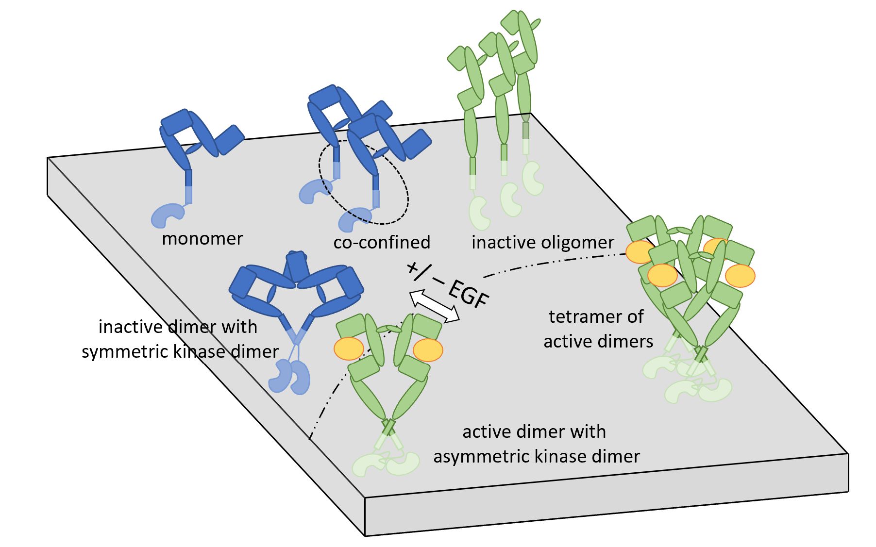 This review explores how receptor tyrosine kinases, especially the ErbB/EGFR receptor family, are regulated by a web of molecular interactions rather than by simple one-to-one binding events. It explains why these receptors are central to controlling cell growth, survival, movement, and fate, and why their dysregulation is so important in cancer. The paper shows how earlier textbook models, such as ligand-induced receptor dimerization, have been refined into more complex views involving preformed clusters, higher-order assemblies, and structural rearrangements. Special attention is given to EGFR, ErbB2/HER2, ErbB3, and ErbB4, whose extracellular, transmembrane, juxtamembrane, kinase, and regulatory domains cooperate to determine signaling output. The authors emphasize that receptor activation is not controlled only by protein–protein contacts, but also by the membrane environment in which these receptors are embedded. Lipids, membrane organization, dipole potential, and lipid–protein interactions can modulate receptor clustering, conformation, and downstream signaling. This membrane-centered perspective helps explain how subtle changes in cancer-cell membranes may contribute to abnormal receptor activity. The review also discusses how modern structural biology, molecular biology, and biophysical fluorescence-based methods have advanced our understanding of receptor tyrosine kinase regulation. By connecting receptor organization, membrane biology, and cancer signaling, the paper highlights new ways to think about therapeutic targeting beyond simply blocking kinase activity. Overall, it presents receptor tyrosine kinases as dynamic, membrane-regulated signaling platforms whose behavior depends on coordinated interactions among ligands, receptors, lipids, and cellular context.
Biophysical
experiments reveal a protective role of protein phosphatase Z1 against
oxidative damage of the cell membrane in Candida albicans
Hajdu T, Szabó K, Jakab Á, Pócsi I, Dombrádi V, Nagy P
Free Rad Biol Med, 176: 222-227. (2021)
Pubmed
DOI
 In a collaborative study, the fungus-specific protein phosphatase Z1 (Ppz1) enzyme has been investigated and it was found to be crucial in the defence against oxidative stress. Researchers in the Department of Biophysics and Cell Biology, Department of Medical Chemistry and Department of Molecular Biotechnology and Microbiology proved that the deletion of the gene of Ppz1 leads to low membrane compactness and an increase in the sensitivity to oxidative challenge in Candida albicans cells. Examining the membrane fluidity-related generalized polarization reveals that while most native cells (WT) expressing Ppz1 were able to neutralize the effect of the applied oxidative agent, tert-butyl-hydroperoxide (tBOOH), mutant cells lacking Ppz1 expression (KO) suffered from latent oxidative damage and were characterized by incompact membrane. The lack of Ppz1 also manifests in increased membrane lipid peroxidation. Investigation of the lateral mobility of lipids revealed that the lack of Ppz1 combined with oxidative stress reduces the lateral diffusion of lipids resulting in reduced competitive fitness. According to these data, applying mild oxidative treatments together with prospective Ppz1 inhibitor molecules may tackle fungal infections caused by drug-resistant Candida albicans species. The study also revealed that changes in membrane characteristics may be used by fungal cells as a cue for responding to changes in the environment.
Statin-boosted cellular uptake and endosomal escape of penetratin due to reduced membrane dipole potential
Batta G, Kárpáti L, Henrique GF, Tóth G, Tarapcsák S, Kovács T, Zákány F, Mándity IM, Nagy P
Br J Pharmacol, 178: 3667-3681. (2021)
Pubmed
DOI
 Research carried out in collaboration between the Department of Biophysics and Cell Biology, and the Department of Genetics of Applied Microbiology at the University of Debrecen, and the Department of Organic Chemistry, Semmelweis University revealed that cellular uptake of cell penetrating peptides (CPP) depends on the membrane dipole potential and that it can be enhanced by reducing the dipole potential. Cell penetrating peptides are molecules capable of traversing the intact cell membrane even when loaded with cargo, e.g. membrane impermeable drugs. The research group of Peter Nagy found that this uptake takes place by endocytosis followed by endosomal escape. Since most cell penetrating peptides are positively charged, the intramembrane, positive dipole potential generated by the ordered arrangement of dipoles in the membrane, inhibits their membrane crossing. Atorvastatin, widely applied in the treatment of high blood cholesterol levels, decreases the membrane dipole potential and consequently enhances the cellular uptake of cell penetrating peptides. The findings lay the foundation for applying the revealed principle for boosting the cellular uptake of drugs in the treatment of human diseases.
An
ω-3, but Not an ω-6 Polyunsaturated Fatty Acid Decreases Membrane
Dipole Potential and Stimulates Endo-Lysosomal Escape of Penetratin
Zákány F, Szabó M, Batta G, Kárpáti L, Mándity M. István, Fülöp P, Varga Z, Panyi G, Nagy P, Kovács T
Frontiers in Cell and Developmental Biology 9: 647300. (2021)
Pubmed
DOI
Epigallocatechin
3-O-gallate induces 67 kDa laminin receptor-mediated cell death
accompanied by downregulation of ErbB proteins and altered lipid raft
clustering in mammary and epidermoid carcinoma cells
Mocanu MM, Ganea C, Georgescu L, Váradi T, Shrestha D, Baran I, Katona E, Nagy P, Szöllősi J.
J Nat Prod. 77(2):250-7 (2014)
Pubmed
DOI